

 Endobronchial ultrasound-guided transbronchial needle aspiration is a valuable diagnostic tool for evaluating abnormalities within the bronchial tree. It allows for targeted sampling of suspicious lesions or endobronchial pathology, aiding in the diagnosis of lung cancer, infections, inflammatory conditions, and other bronchial abnormalities. The procedure is typically performed by a pulmonologist or interventional bronchoscopist who has specialized training and expertise in bronchoscopy and biopsy techniques.
Endobronchial ultrasound-guided transbronchial needle aspiration (EBUS-TBNA) is a specialized procedure that combines bronchoscopy and ultrasound technology to obtain tissue samples from structures adjacent to the airways, such as lymph nodes or masses. It is primarily used for the diagnosis and staging of lung cancer and other thoracic conditions.
Here's an overview of the process of endobronchial ultrasound-guided transbronchial needle aspiration (EBUS-TBNA):
1. Preparation: Before the procedure, the patient may receive local anesthesia to numb the throat and reduce discomfort. Conscious sedation or general anesthesia may also be administered to help the patient relax or perform more extensive interventions.
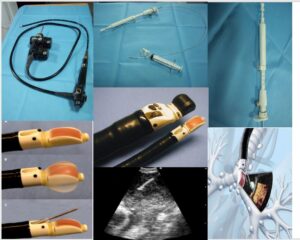
2. Insertion of the EBUS bronchoscope: The EBUS bronchoscope, which incorporates an ultrasound probe at its tip, is inserted through the nose or mouth and guided into the airways. The bronchoscopist navigates the bronchoscope to the target area using real-time ultrasound imaging.
3. Visualization and target identification: The ultrasound imaging allows the bronchoscopist to visualize nearby structures, such as lymph nodes, tumors, or abnormal masses. This enables precise targeting and identification of the areas for sampling.
4. Needle aspiration: A specialized needle, called a transbronchial needle, is passed through the working channel of the EBUS bronchoscope. Under ultrasound guidance, the needle is advanced through the airway wall and into the target structure, such as a lymph node. The needle is then used to obtain tissue samples by applying suction and moving the needle back and forth within the target area.
5. Specimen collection and processing: The collected tissue samples are sent to a pathology laboratory for processing and analysis. The samples are stained and examined under a microscope by a pathologist specializing in lung tissue analysis. The pathologist evaluates the samples to determine the nature of the tissue, identify any abnormalities, and establish a diagnosis or stage the disease.
6. Post-procedure care: After the procedure, the patient is monitored for any immediate complications, such as bleeding or respiratory distress. The patient may experience a sore throat, cough, or mild bleeding, which typically resolve within a short time. Recovery time is generally minimal, and patients can often resume normal activities shortly after the procedure.
EBUS-TBNA is a valuable technique for diagnosing and staging lung cancer, as well as evaluating other thoracic conditions. It provides real-time imaging guidance to obtain tissue samples from structures adjacent to the airways, offering a minimally invasive alternative to more invasive surgical procedures. The procedure is typically performed by a pulmonologist or interventional bronchoscopist who has specialized training and expertise in EBUS-TBNA techniques.